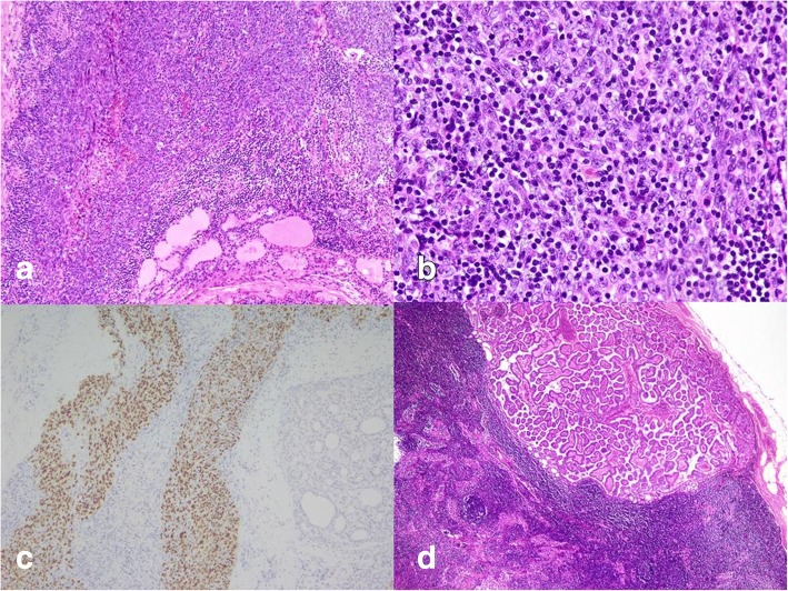
Fig. 3.
Histopathological findings of left thyroid tumor: sheets or nests of epithelial cells infiltrated by lymphoplasmacytic cells (a. H&E, 100X; b. H&E, 200×); malignant epithelial cells positive for EBER ISH (c. Hematoxylin counterstain, 200×); metastatic papillary carcinoma found in right neck lymph node (d. H&E, 40×)

