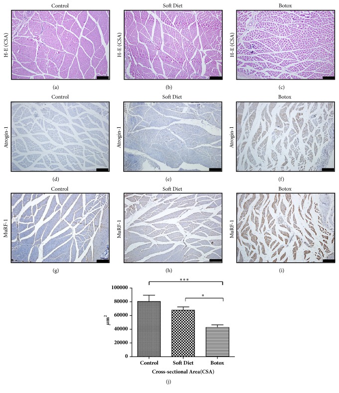
Figure 1.
Histological changes and atrogin-1/MuRF-1 immunohistochemical staining of masseter muscles in the control, SD, and BTX groups. After 4 weeks of treatment, H-E staining of masseter muscle revealed an obvious atrophic condition in the BTX group, whereas no difference in the SD group was seen (a–c, j). The distinctive expression of atrogin-1/MuRF-1 could be easily found in the BTX group compared to that in the control and SD groups (d–i) (∗p<0.05;∗∗p<0.01;∗∗∗p<0.001). Bar=100 μm.

