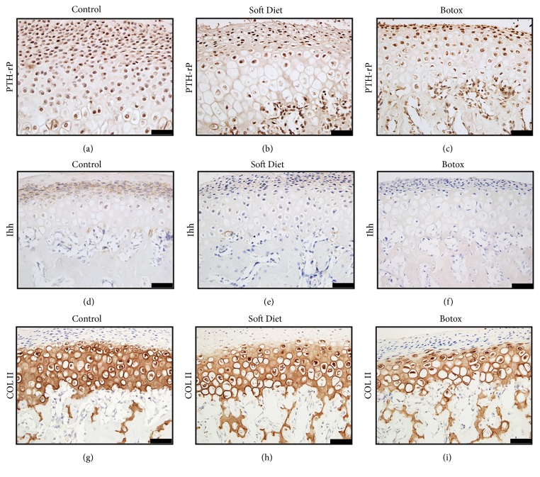
Figure 4.
Immunostaining changes of condylar cartilage. The PTHrP positive cells were distributed in the proliferative and early hypertrophic layers (a–c). Ihh was expressed in the extracellular space and concentrated within the proliferative layer (d–f). The Col2a1 positive area was shown in the hyperplastic layers; the intensity and area predicted the matrix's volume (g–i) (∗p<0.05;∗∗p<0.01;∗∗∗p<0.001). Bar=25 μm.

