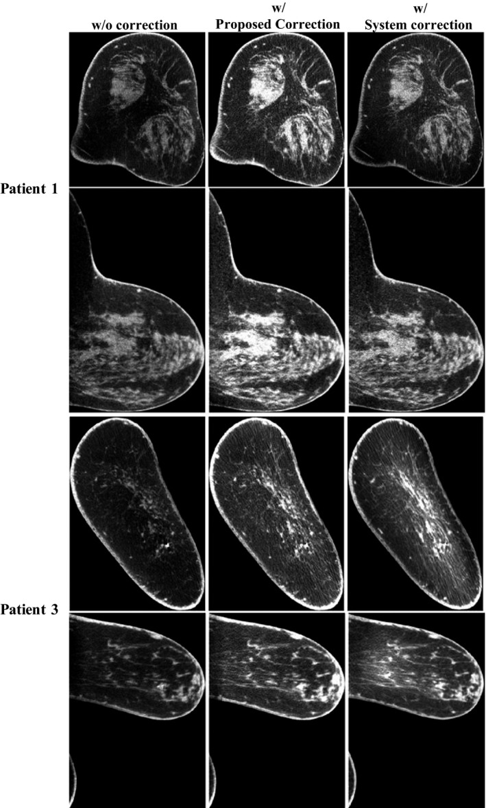
Figure 6.

Comparison of the uncorrected image, the corrected image with the proposed scatter correction method and the corrected image with the system‐embedded software. The images are taken on Patient 1 and 3, but at slices different from those shown in Fig. 2. Display window: [0.2 0.3] cm−1.
