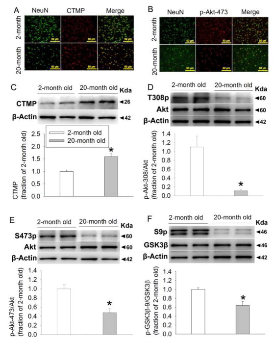
FIGURE 1. Aging increases CTMP and decreases phosphorylated Akt and GSK-3β in the cerebral cortex of Fischer 344 rats.
(A) Representative images of immunolabeling of NeuN (green) and CTMP (red). Magnification x 200. Scale bar equals to 50 μm. (B) Representative images of immunolabeling of NeuN (green) and Akt phosphorylated at Ser473 (p-Ser473-Akt) (red). Magnification x 200. Scale bar equals to 50 μm. (C) Representative images and quantification of Western blot of CTMP. (D) Representative images and quantification of Western blot of Akt phosphorylated at Thr308. (E) Representative images and quantification of Western blot of Akt phosphorylated at Ser473. (F) Representative images and quantification of Western blot of GSK-3β phosphorylated at Ser9. Data are mean ± S.E.M. (n = 6–9). * P < 0.05 compared with 2-month old rats.

