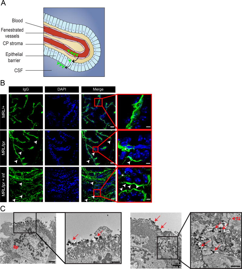
Figure 3. BCSFB dysfunction in NPSLE mice points to an alternative route of antibody entry into the CNS.
Examination of endogenous IgG with confocal and TEM imaging indicates abnormal localization of antibodies at the ventricular side of the BCSFB. A. Illustration of the choroid plexus (CP), located in brain ventricles. Tissue structure consists of permeable vessels in the middle and an epithelial barrier at the perimeter of the CP. Antibodies (depicted in green) demarcating the possible route from the blood through fenestrated vessels into the CP stroma and eventually to the apical side of the barrier (modified from9). B. Confocal images of 10 µm sagittal brain sections of 16 week old MRL/lpr mice stained for endogenous IgG (green) and nuclei (DAPI, blue). As expected, IgG is found in CP vessels (asterisks). IgG is also found at the apical side of the epithelial barrier only in 16 week old MRL/lpr mice (lower panels, arrow heads) but not in control MRL/+ mice (upper panel). Ventricular BCSFB IgG staining is more abundant in CP regions where immune cells infiltrated the stroma (highly infiltrated regions, lower panel. less infiltrated regions, middle panel). Scale bars 20 µm and 5 µm in insets. C. TEM imaging of 16 week old MRL/lpr CP stained with IgG-HRP. IgG is localized at the ventricular surface of the BCSFB, covering epithelial cells microvilli (left panel, arrows in inset). Abundant IgG deposits are also found in the basolateral space between epithelial cells (right panel, arrows), reaching the epithelial tight junction (right panel, arrow head in inset). Scale bars 2 µm, and 1 µm in insets.

