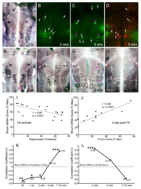
Fig. 2. RhoA mRNA expression in reticulospinal neurons is negatively associated with regenerative probability.
At 1 week post-SCI, RhoA mRNA levels (E) were similar to those in normal brain (A) and negatively correlated with regenerative probability of reticulospinal neurons (I and K) during the first two weeks. The magenta arrowhead in E points to a retracting axon tip labeled for RhoA mRNA. After 2 weeks post-SCI, apoptotic signaling was detected in reticulospinal neurons by FLICA (B–D), followed by wholemount ISH for RhoA (F–H). At 2 weeks post-transection, RhoA mRNA expression was seen selectively in caspase-positive RNs (white arrows in B & F; J). At 4 weeks post-transection (C, G), expression of RhoA mRNA in FLICA+ neurons varied, low in some (magenta arrows), high or unchanged in others (white arrows). At 8 weeks post-transection (D, H), regenerated reticulospinal neurons were labeled with DTMR applied 5 mm caudal to the original transection. One week later, RhoA mRNA expression was normal in regenerated reticulospinal neurons (H, white arrowheads) but reduced in all FLICA+ cells (white arrows). The correlation coefficients (r) between RhoA mRNA score and either regenerative probability of reticulospinal neurons or FLICA intensity score at different post-transection times are plotted in K & L. *** p ≤ 0.001; ** p ≤ 0.01; * p ≤ 0.05; wk = week; Mth, Mauthner neuron.

