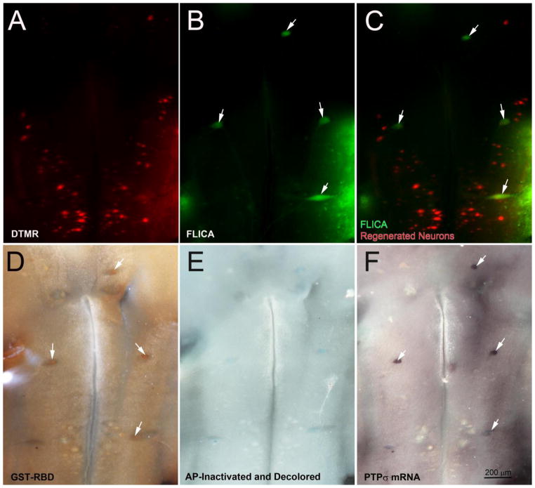
Fig. 8. Co-localization of activated caspases and RhoA with PTPσ mRNA.
At 8 weeks post-SCI, reticulospinal neurons with regenerated axons were labeled by DTMR applied to a 2nd transection 5 mm caudal to the original lesion. After allowing one more week for retrodrade labeling, brains were removed and processed as indicated. Neurons whose axons had regenerated are labeled red (A). There was no overlap with FLICA-positive neurons (B, C). Lamprey brains were further processed for activated RhoA protein (D) followed by ISH for PTPσ (F), a receptor for CSPGs. White arrows point to neurons that were triply labeled by FLICA, GST-RBD and PTPσ ISH.

