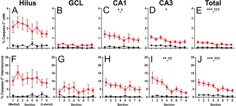
Figure 2.

Medial to lateral gradient of activated caspase-3 positive cells and interneurons in the hippocampus at P7. (A-E) The percent of activated caspase-3 positive cells as a percentage of total DAPI nuclei in every medial (section 1) to lateral (section 8) section in the hilus (A), GCL (B), CA1 (C), CA3 (D), and combined hippocampal regions (E). (F-J) The percent of activated caspase-3 positive interneurons as a function of total Venus- + cells in every medial to lateral section in the hilus (F), GCL (G), CA1 (H), CA3 (I), and combined hippocampal regions (J). Black circles represent air-exposed mice, red squares represent EtOH-exposed mice. Asterisk(*) denotes a significant within-subjects effect of medial to lateral tissue section on the percentage of either activated caspase-3 positive cells or activated caspase-3 positive interneurons at p < 0.05, ** p < 0.01, *** p < 0.001. Dagger(†) denotes a significant interaction of medial to lateral tissue section and vapor chamber exposure condition on the percentage of activated caspase-3 positive cells at p < 0.05, †† p < 0.01, ††† p < 0.001. n = 5 mice per vapor chamber exposure condition.
