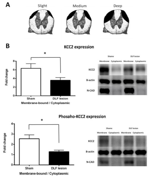
Fig. 4.
DLF lesions down-regulate KCC2 expression. (A) Samples were selected to illustrate the range of DLF lesions, from deep through medium to slight (n=4, 6, and 3, respectively). (B) The fold change in the membrane-bound/cytoplasmic KCC2 and phosphor-KCC2 ratio in DLF lesioned (black) and sham-operated (white) rats. Representative Western blots are provided to the right of each panel. The error bars depict ± SEM.

