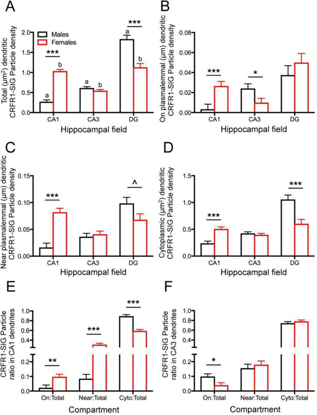
Fig. 2. Sex differences in distribution of CRFR1-SIG particles in CA1, CA3, and DG in the unstressed control groups.

A. In control rats, the total density of CRFR1-SIG particles in dendrites (# SIG/μm2) was significantly lower in CA1 and higher in the DG of males compared to females. Moreover, significantly lower densities of CRFR1-SIG particles in dendrites were seen in CA1 compared to CA3 and DG in males and higher densities of CRFR1 SIG particles in CA1 and DG compared to CA3 in females. B. The density of CRFR1 SIG particles on the plasma membrane (# SIG/μm) in dendrites was significantly lower in CA1 and higher in CA3 of males compared to females. C. The density of CRFR1-SIG particles near the plasma membrane (# SIG/μm) in dendrites was significantly lower in CA1 and higher in DG of males compared to females. D. The density of CRFR1 SIG particles in the cytoplasm (# SIG/μm2) of dendrites was significantly lower in CA1 and higher in DG of males compared to females. E. In CA1, a smaller partitioning ratio of CRFR1-SIG particles was on the plasma membrane (On/Total) and near the plasma membrane (Near/Total), and a greater ratio of cytoplasmic CRFR1-SIG particles (Cyto/Total) in males compared to females. F. In CA3, males compared to females had a greater ratio of CRFR1-SIG particles on the plasma membrane (PM/total). No significant differences in partitioning ratio were observed in hilar interneurons. ***p < 0.001; **p < 0.01; *p < 0.05; ^p = 0.052. a p < 0.001 (CA1 vs. DG; CA3 vs. DG) and p < 0.01 (CA1 vs. CA3); b p < 0.001 (CA1 vs. CA3; CA3 vs. DG). N = 3 rats/group; n = 50 dendrites/rat/area.
