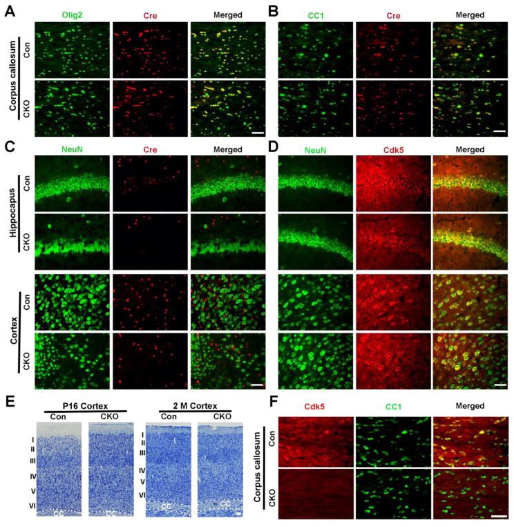
Figure 2. CNP-targeted deletion of Cdk5 is restricted to oligodendrocyte lineages in CNP;Cdk5 CKO mice.
(A–B) Double immunostaining showed co-localization of Cre and olig2+ (A) and CC1+ (B) in the corpus callosum of 2-month-old CNP;Cdk5 CKO and controls. (C) Lack of Cre expression in either hippocampal or cortical neurons identified by NeuN in CNP;Cdk5 CKO mice and controls. (D) Cdk5 expression presents in NeuN+ cells in the cortex and hippocampus of CNP;Cdk5 CKO mice. (E) Representative Nissl-stained images show relatively normal cortical lamination in CNP;Cdk5 CKO at P16 and 2-months of age. (F) Lack of Cdk5 expression in CC+ cells in the corpus callosum of 2-month-old CNP;Cdk5 CKO mice. Scale bars = 50 μm.

