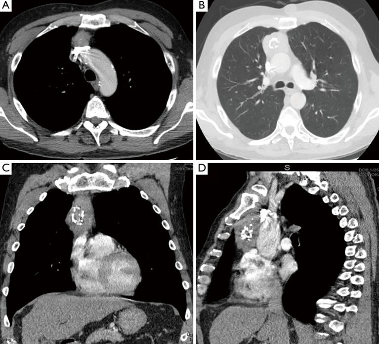
Figure 1.
Chest CT-scan images. (A) Horizontal section: the superior portion of the thymic mass is lying on the left innominate vein; (B) horizontal section: the largest transverse diameter portion of the thymic mass is shown with the relationship of ascending aorta; (C) coronal section: the ovoid-elongated mass is located on the right side of the median line; (D) sagittal section: the boundaries of the thymic mass are shown between the sternum and heart. CT, computed tomography.

