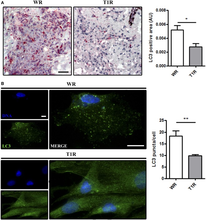
Figure 3.
Increase of autophagy levels in skin lesions of multibacillary patients that did not develop reversal reactional episodes (WR). Skin lesion samples were obtained from multibacillary patients who developed (T1R) or not (WR) reversal reactional episodes and analyzed as indicated. (A,B) Increased LC3 expression in skin lesion cells of WR patients. (A) Immunohistochemical (IHC) analysis of endogenous LC3. Representative micrographs from WR (n = 3) and T1R (n = 4) patients are shown. IHC images were quantified and data are expressed as arbitrary units (AU). Bars represent the mean values ± SEM. *p < 0.05. Scale bar: 50 µm. (B) Macrophages were isolated from skin lesions of multibacillary patients who developed (T1R) or not (WR) episodes of reversal reaction in the future, and cultured for 18 h. Cells were fixed and stained with the anti-LC3 antibody (green) and DAPI (blue). Macrophages of WR skin lesions showed enhanced LC3 puncta formation as compared to T1R macrophages. Immunofluorescence images were quantified and bars represent the mean values of the number of LC3 puncta per cell ± SEM (WR, n = 3; T1R, n = 3) (**p < 0.01). Scale bar: 20 µm.

