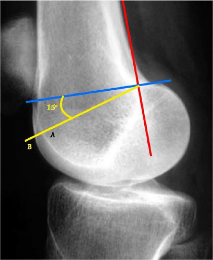
Fig. 4.

The trochlear depth measurement is performed on a true lateral radiograph view. A tangent to the posterior femoral cortex (red line) and a perpendicular line at the most proximal part of the posterior condyles (blue line) are drawn. A (yellow) line subtended 15° from the perpendicular line is now used to measure the trochlea depth (AB length).
