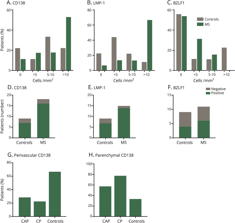
Figure 5. Increased frequency of parenchymal CD138- and LMP-1–positive cells in MS.
Formalin-fixed paraffin embedded brain tissue from MS and control brains without neurologic disease were cut into 4-μm sections. Hematoxylin and eosin (H&E) and immunohistochemistry were performed using antibodies against latent membrane protein 1 (LMP-1), Epstein-Barr virus (EBV) immediate-early lytic gene (BZLF1), and Syndecan-1 (CD138), a plasma cell marker. For each MS and control sample, the number of CD138+, LMP-1+, and BZLF1+ cells with a visible nucleus was counted manually to allow semiquantitative analysis and categorization of these markers. Results are semiquantitative and expressed as percentage of patients expressing as no cells/mm2, <5 cells/mm2, 5–10 cells/mm2, and >10 cells/mm2 (A–C). Semiquantitative analysis of CD138 (D) and EBV antigen-positive cells (E,F) in MS and healthy control samples (D-F). CD138+ cells in MS and control samples were characterized by their location in perivascular regions or in the parenchyma (G and H), revealing an increased frequency of parenchymal CD138+ cells in CAPs and CPs vs controls (H). The number of cells was counted from samples (MS: n = 11 autopsy samples and n = 6 biopsy samples; controls samples: n = 9 autopsy samples n = 0 biopsy samples). The number of cells was counted on 3 × 3-cm autopsy sections and on 2 × 2-cm sections for biopsy samples.

