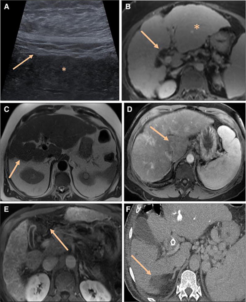
Fig. 1.

a–f Morphologic imaging features of cirrhosis in 6 patients. A) Ultrasound shows a nodular surface (arrow) and coarsened echotexture (*). MRI images of cirrhotic livers show B) a nodular surface contour, hypertrophy of lateral left lobe (*), and expanded hilar periportal space (arrow) on post contrast T1 FS sequence, C) atrophic right hepatic lobe (arrow) on axial T2 Half Fourier Acquisition Single Shot Turbo Spin Echo (HASTE) sequence, D) hypertrophy of caudate lobe on post contrast T1 FS sequence (arrow), E) expanded gallbladder fossa and atrophic medial segment left lobe on post contrast T1 FS sequence (arrow). F) Postcontrast CT in the portal venous phase shows a right hepatic posterior “notch” (arrow)
