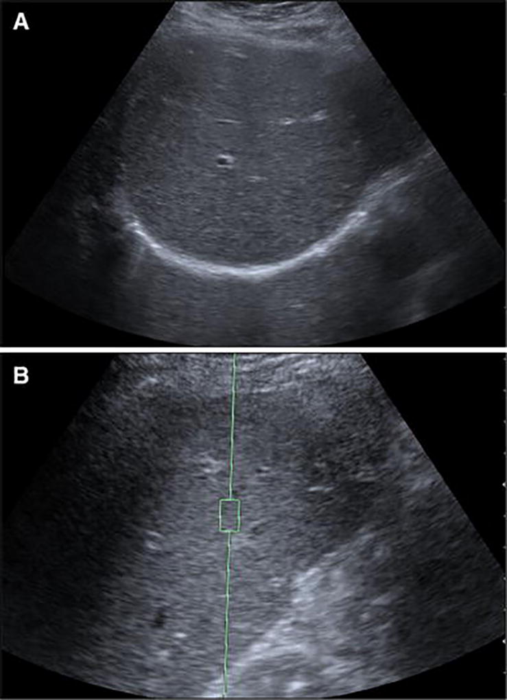
Fig. 2.

a–b Ultrasound of a 61-year-old woman with HIV and hepatitis C presenting for fibrosis screening shows potentially treatable liver fibrosis on ARFI prior to morphologic changes on grayscale ultrasound. A) Grayscale ultrasound of the right hepatic lobe shows a normal smooth echotexture. B) ARFI shows a shear wave velocity of 1.79 m/s (F3)
