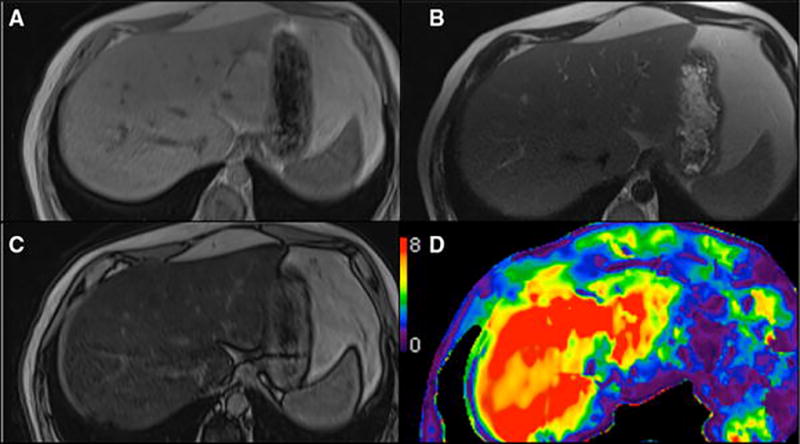
Fig. 4.

a–d 57-year-old man with fatty liver disease. Hepatic steatosis is seen with signal loss on the opposed phase imaging relative to the in phase images (A and B), but with normal liver morphology on T1 and T2-weighted imaging (A–C). D) MR elastography shows unsuspected cirrhosis (stiffness 7.5 kPa), making biopsy unnecessary
