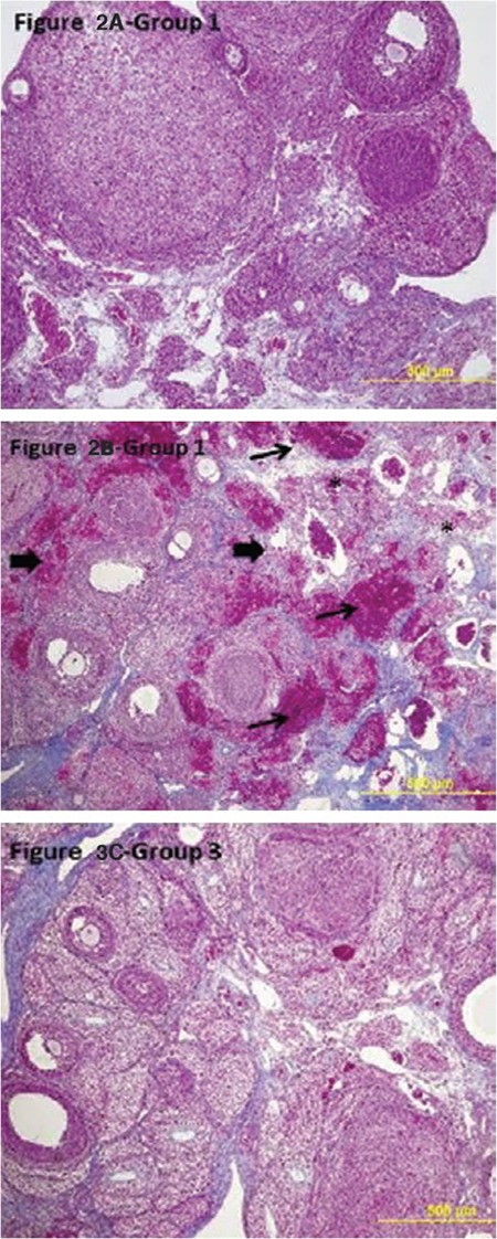
Figure 2. Light microscopic findings of the groups (100x, Original magnification, Masson trichrome). Ovarian sections in group 2 (I/R) showed severe damage, infiltration of polymorphonuclear leukocytes, vascular congestion, interstitial edema*, hemorrhage → → I/R: Ischemia/reperfusion.

