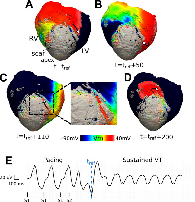
Figure 5.

Reentry involving a sub-epicardial channel of viable tissue in heart 5 (CL = 230 ms). (A-D) Transmembrane voltage maps showing one cycle of reentry. The wave traverses the channel in (B) and (C); its exit from the channel is followed by a centrifugal activation of the tissue outside the scar in (D). The length of the epicardial channel is approximately 30 mm and the VT pathlength is 74 mm. A border layer around the scar has been rendered semi-transparent to visualize channel position. (E) Pseudo-ECG. tref : reference time.
