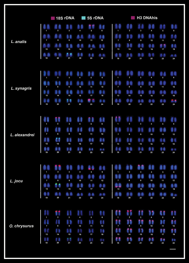
Figure 1.
Fluorescence in situ hybridization (FISH) in the metaphase chromosomes of snappers species highlighting the chromosomal location of 18S rDNA (red) and 5S rDNA (green) sites (left column) and of hisDNA H3 (red) sites (right column). Chromosomes were counterstained with DAPI (blue). Bar = 5 µm.

