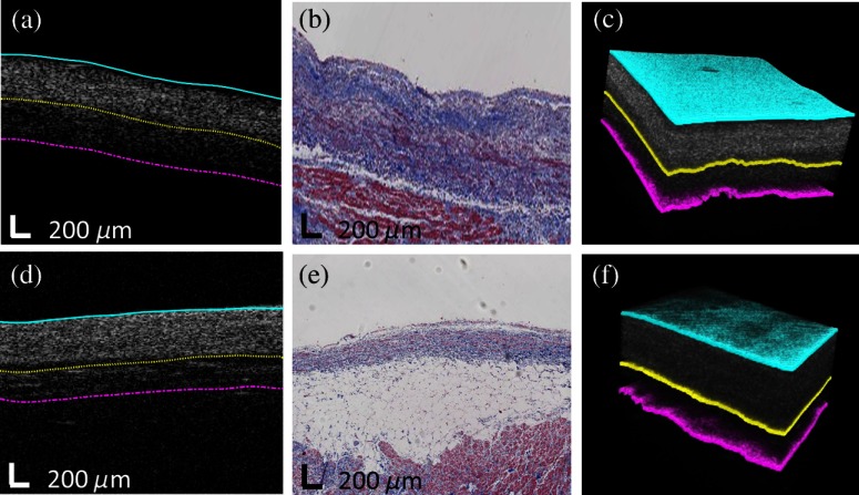
Fig. 4.
Segmentation results from human atria. (a) and (d) Original OCT images overlaid with automated segmentation result; (b) and (e) corresponding trichrome histology image; (c) and (e) 3-D segmentation results. The automated results in both 2-D and 3-D show great agreement with histology images.

