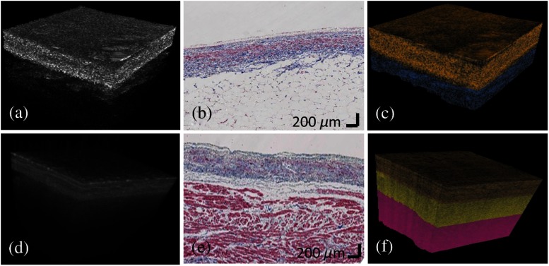
Fig. 8.
3-D classification results from human atria. (a) and (d) Original OCT volumes; (b) and (e) histology images; (c) and (f) color-coded classification results. Gold, yellow, red, and blue colors represent dense collagen, loose collagen, normal myocardium, and adipose tissue, respectively. The classification results delineated the layer structure and agreed with trichrome histology.

