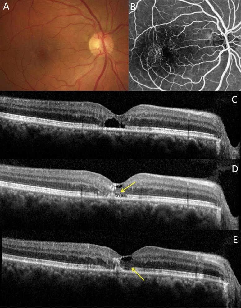
Figure 8.
Retinal response to NRT in MacTel patient. (A) Baseline fundus color image. (B) Baseline fluorescein angiography with typical telangiectasic vessels temporal to fovea. (C) Baseline OCT image with subfoveal cavitation and loss of IS/OS junction line integrity. (D) Partial restoration of outer nuclear layer and external limiting membrane (arrow) at 5 months; (E) at 12 months, the IS/OS junction line became more visible (arrow).

