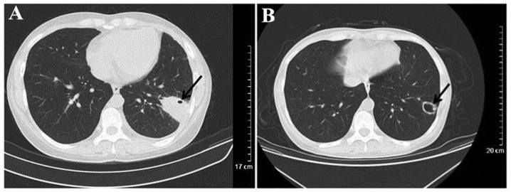
Figure 7.
Computed tomography scanning of a patient with WG misdiagnosed as infectious lesions. (A) Irregular consolidation in the substrate out of left lower lung, and punctiform vesicle in the consolidation (black arrow). (B) At 3 months post predisone treatment, the lesion markedly reduced in size and evolved into a cavity with a thin-wall (black arrow).

