Figure 1.

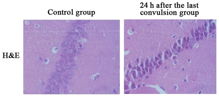
H&E staining displays changes of CA1 apoptosis of hippocampus during developmental stage in control group and 24 h after the last convulsion group (×400 magnification).

H&E staining displays changes of CA1 apoptosis of hippocampus during developmental stage in control group and 24 h after the last convulsion group (×400 magnification).