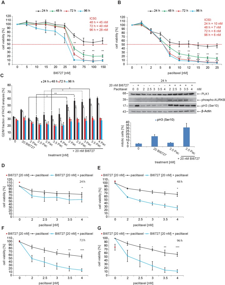
Figure 1. BI6727 treatment sensitizes ovarian cancer cells to paclitaxel.
(A) OVCAR-3 cells were treated with increasing concentrations of BI6727 or (B) of paclitaxel (Pac). Cell viability was measured over 4 d using the Cell Titer-Blue® Cell Viability Assay. (C) (Left panel) The G2/M fraction was determined over 4 d post-treatment using flow cytometry. Measurements were statistically significant by two-tailed Student's t-test (*P ≤ 0.05; **P ≤ 0.01). Each bar graph represents the mean value ± SEM (n=3). (Upper right panel) Endogenous levels of PLK1, Cyclin B1, phospho-Histone H3 and phospho-Aurora B were determined by immunoblotting. β-Actin served as loading control. (Lower right panel) The mitotic index was determined by measuring pH3(Ser10) levels. (D-G) OVCAR-3 cells were treated with either 20 nM BI6727 or increasing paclitaxel concentrations or both for 4 d. The cell viability was determined using the Cell Titer-Blue® Cell Viability Assay. Measurements were statistically significant by two-tailed Student's t-test (*P ≤ 0.05; **P ≤ 0.01; ***P ≤ 0.001). Each measurement represents the mean value ± SEM (n=3).

