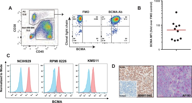
Figure 1. BCMA expression on MM patient samples.
(A) An example of flow cytometry analysis of a bone marrow (BM) sample from an MM patient is shown. Plasma cells (PC; both malignant and non-malignant) were identified by gating bone marrow on live single cells followed by using CD38 and CD45 staining as shown. Malignant PCs were evaluated for BCMA expression with fluorescence minus one, FMO, used as control. (B) Ten consecutive MM patients were analyzed for BCMA expression as in Figure 1A. BCMA median expression was 86% (red line). (C) BCMA expression (dark histogram) on 3 established multiple myeloma cell lines assessed by flow cytometer. The lighter histogram shows staining of a matched isotype control. (D) Representative immunohistochemical staining of a bone marrow biopsy specimen stained for BCMA using two commercially available antibodies, B0807-50G (brown staining, primaryantibody concentration of 0.6 μg/ml, US Biological) and AF193 (magenta staining, primary antibody concentration of 2.0 μgml, R&D Systems).

