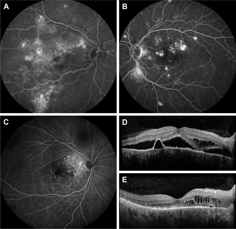
Figure 1.
Illustration of the 4 criteria of severity on FA and OCT.
Notes: The 4 criteria are as follows: 1) cumulative areas of >5 DD of diffuse atrophic retinal pigment epithelium alterations as visualized on mid-phase FA (A). 2) Multiple (at least 2) “hot spots” of leakage separated by at least 1 DD of non-hyperfluorescent healthy-appearing retina on mid-phase FA (B). 3) An area of diffuse fluorescein leakage with a surface of >1 DD on mid-phase FA, without an evident leaking focus (C). 4) Presence of posterior cystoid retinal degeneration on OCT (D and E).
Abbreviations: DD, optic disc diameters; FA, fluorescein angiography; OCT, optical coherence tomography.

