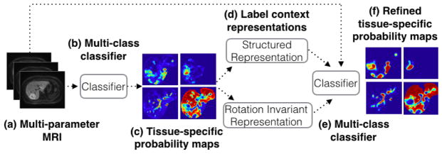
Fig. 2.
Representation of the unique iterative classification method: (a) multi-parameter MRI input images, (b) multi-class classifier, in our case a random forest, (c) tissue-specific probability maps output by the classifier, (d) structured and rotationally-invariant label context representations are computed and used as input to another classifier, in addition to the original multi-parameter MRI imaging, (f) tissue-specific probability maps output by the classifier, and the entire process is iterated.

