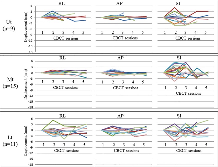
Fig 4. Plots of inter-CBCT session marker displacement using breath-hold.
This figure provides plots of inter-CBCT session marker displacement using breath-hold. CBCT data based on the first CBCT scan are shown. Three, four, and five CBCT scans were performed in 7, 4, and 5 patients, respectively. The different colors stand for the data from the different metal markers. CBCT: Cone beam computed tomography; Ut: upper thoracic esophagus; Mt: middle thoracic esophagus; Lt: lower thoracic esophagus /esophagogastric junction; RL: right–left; AP: anterior–posterior; SI: superior-inferior.

