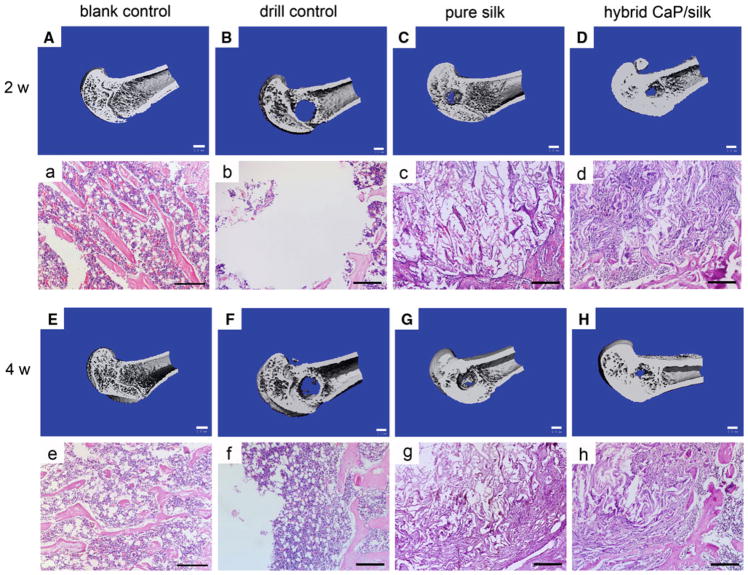
Fig. 4.
3D reconstruction of distal femur represented mineralized bone formation within defects in the blank control group, drill control group, pure silk scaffolds group and the hybrid CaP/silk scaffolds group (A–H; bar = 1 mm). H&E staining revealed new bone matrix deposition within defects in the above mentioned groups (a–h; bar = 200 μm)

