Figure 6.
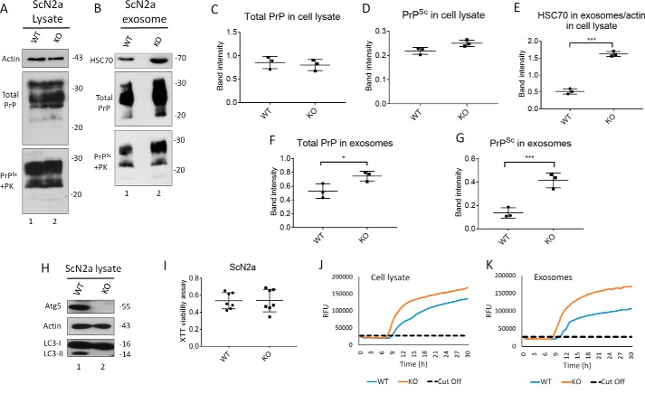
Knockout of Atg5 increases exosome release and exosomal PrPSc in ScN2a cells. A and B, Western blotting of cell lysate and exosomes from ScN2a cells respectively, either WT or KO for Atg5. HSC70 was used as exosomal markers. Total PrP and PrPSc were detected using mAb 4H11 antibody. C and D, densitometric analysis for either total PrP or PrPSc, respectively, from WT or KO ScN2a cell lysate normalized with actin (± S.D.; n = 3 experiments). E, densitometric analysis for exosomal HSC70 normalized with actin in the corresponding cell lysate (± S.D.; n = 3 experiments). ***, p < 0.001. F and G, densitometric analysis for either total PrP or PrPSc, respectively, from WT or KO ScN2a exosomes normalized with actin in the corresponding cell lysate (± S.D.; n = 3 experiments). ***, p < 0.001; *, p < 0.05. H, immunoblot showing complete knockout of Atg5 compared with WT ScN2a cells. Actin was used as loading control. Atg5 KO resulted in complete absence of LC3-II, confirming disruption of autophagy machinery. I, XTT viability assay. (OD = 490 nm). The cells were cultured for 48 h for XTT viability assay (n = 7 replicates). J and K, RT-QuIC for WT or KO ScN2a cells.

