Figure 7.
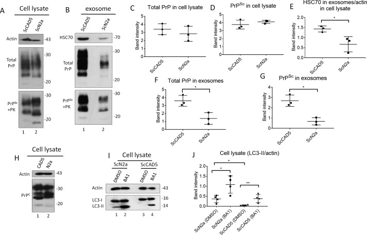
ScCAD5 cells release more exosomes and PrPSc compared with ScN2a cells, which is inversely correlated to autophagy competence. Comparable numbers of ScN2a and ScCAD5 cells were used in this experiment. A and B, cell lysate and exosomes isolated from conditioned media from ScCAD5 and ScN2a cells were analyzed in immunoblot for HSC70 and PrP. C and D, densitometric analysis for either total PrP or PrPSc respectively from ScCAD5 or ScN2a cell lysate normalized with actin (± S.D.; n = 3 experiments). E, densitometric analysis for exosomal HSC70 normalized with actin in the corresponding cell lysate (± S.D.; n = 3 experiments). *, p < 0.05. F and G, densitometric analysis for either total PrP or PrPSc, respectively, from ScCAD5 or ScN2a exosomes normalized with actin in the corresponding cell lysate (± S.D.; n = 3 experiments). *, p < 0.05. H, immunoblot comparing PrPC levels between CAD5 and N2a cells using mAb 4H11. I, immunoblot comparing the level of LC3-I to LC3-II between ScN2a and ScCAD5 cells with and without treatment with bafilomycin A1 (BA1) for 4 h. Actin was used as a loading control. J, densitometric analysis for LC3-II protein levels normalized with actin (± S.D.; n = 4 replicates). *, p < 0.05, **, p < 0.01.

