Figure 1.
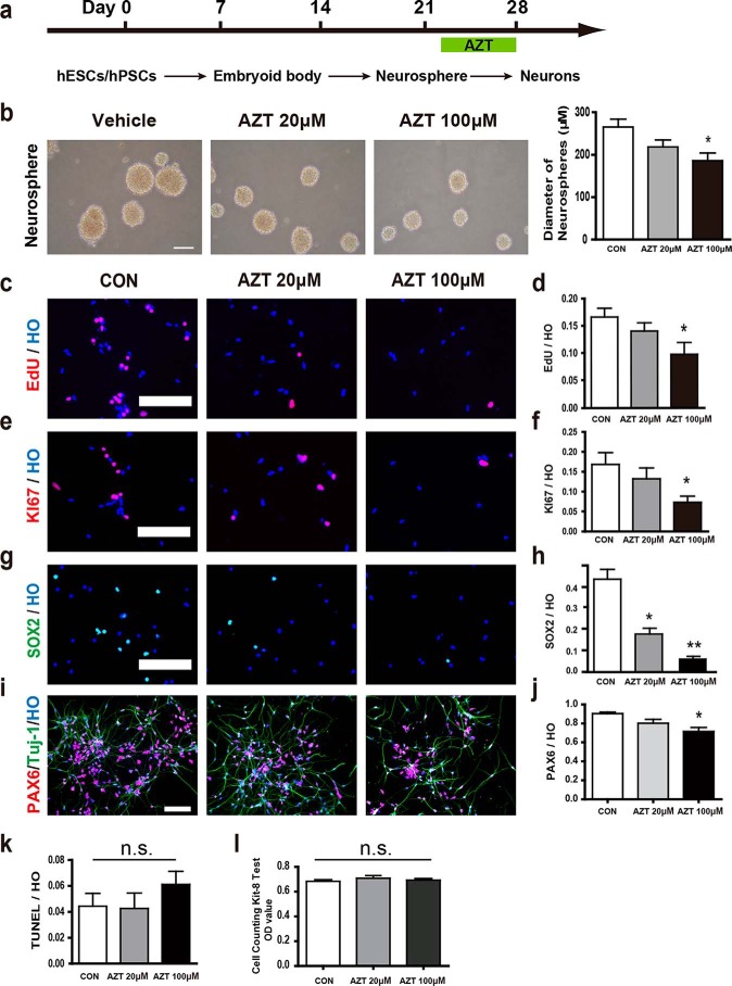
AZT inhibited the proliferation of hPSC-derived neural progenitors. a, brief time line of human stem cell differentiation for generating cortical neurons. b, brightfield images of neurospheres. 100 μm AZT treatment significantly reduced the diameter of neurospheres. Scale bar, 250 μm. c and d, EdU incorporation in adherent cells at day 28. 100 μm AZT treatment significantly reduced the percentage of EdU-incorporated cells (EdU/Hoechst). e and f, immunostaining of adherent cells at day 28 for KI67. 100 μm AZT treatment significantly reduced the percentage of KI67-positive cells (KI67/Hoechst). g and h, immunostaining of adherent cells at days 28 and 35 for SOX2. 20 and 100 μm AZT treatments significantly reduced the percentage of SOX2-positive cells (SOX2/Hoechst) at day 28. AZT treatment abolished SOX2 expression in cells at day 35. i and j, immunostaining of adherent cells at day 28 for PAX6. 100 μm AZT treatment significantly reduced the percentage of PAX6-positive cells (PAX6/Hoechst). k, labeling of adherent cells at day 28 for TUNEL. Neither 20 nor 100 μm AZT treatment increased the percentage of TUNEL-positive cells (TUNEL/Hoechst). l, CCK-8 assay of adherent cells at day 28. Neither 20 nor 100 μm AZT treatment increased the OD value of CCK-8 test. Control, n = 5; 20 μm AZT, n = 5; 100 μm AZT, n = 6. *, p < 0.05; **, p < 0.01; n.s., not significant. Scale bars, 100 μm. CON represents control. HO represents Hoechst. Error bars represent S.E.

