Figure 5.

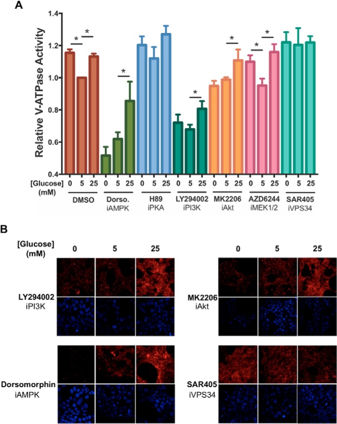
Effect of inhibitors of AMPK, PKA, PI3K, Akt, ERK, or Vps34 on starvation-dependent increases in V-ATPase-dependent lysosomal acidification in HEK293T cells. A, HEK293T cells were loaded with FITC-dextran and maintained in serum-free DMEM containing 5 mm glucose for ∼6 h and then treated with serum-free DMEM containing 0 mm glucose (10 min), 5 mm glucose (1 h), or 25 mm glucose (1 h). Where present, the cells were incubated with indicated inhibitors for 1 h prior to cell homogenization. Inhibitors included dorsomorphin (5 μm, an AMPK inhibitor), H89 (50 μm, a PKA inhibitor), LY294002 (50 μm, a PI3K inhibitor), MK2206 (1 μm, an Akt inhibitor), AZD6244 (1 μm, a MEK1/2 inhibitor), and SAR405 (10 μm, a Vps34 inhibitor). After treatment, cells were homogenized, and V-ATPase-dependent lysosomal acidification was tested as described in Fig. 2A. The error bars represent standard error. The asterisks represent statistically significant differences with a p value less than 0.05 (n ≥ 3). B, HEK293T cells were maintained in serum-free DMEM containing 5 mm glucose for ∼6 h and then treated with serum-free DMEM containing 0 mm glucose (10 min.), 5 mm glucose (1 h), or 25 mm glucose (1 h) in the presence or absence of various signaling pathway inhibitors. Inhibitors were added at the concentrations indicated in A and, where present, were incubated with cells for a total of 1 h. 10 min prior to cell collection, cells were incubated with LysoTracker to stain for acidic intracellular compartments and then fixed as described under “Experimental procedures.” Staining was detected by confocal fluorescence microscopy (red, LysoTracker; blue, DAPI). Drug-free and concanamycin A-treated controls for the experiment are shown in Fig. 2B. Representative images are shown, n = 2.
