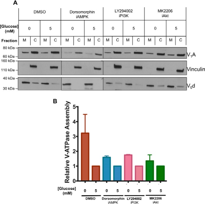
Figure 6.

Effect of inhibitors of AMPK, PI3K, and Akt on starvation-dependent increase in V-ATPase assembly in HEK293T cells. A, HEK293T cells were maintained in serum-free DMEM containing 5 mm glucose for ∼6 h and then treated with serum-free DMEM containing 0 mm glucose (10 min) or 5 mm glucose (10 min) in the presence or absence of the indicated signaling pathway inhibitors. Inhibitors were present at the concentrations indicated in Fig. 5 and, where present, were incubated with cells for 1 h prior to analysis of assembly. Following treatment, assembly was assayed as described in Fig. 1A. Representative Western blots are shown. M indicates membrane and C indicates cytosolic fractions. B, Western blotting quantitation was performed as described in Fig. 1B, with values expressed relative to assembly measured at 5 mm glucose under each set of conditions. Error bars represent standard error, n = 2.
