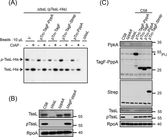
Figure 3.

Both TagF and PppA domains repress T6SS activity independently of the PpkA-mediated TssL phosphorylation pathway in A. tumefaciens. A, Phos-tag SDS-PAGE analysis to detect the phosphorylation status of TssL-His. Shown is Western blot analysis of the same volumes of Ni-NTA resins (10 μl) associated with TssL-His from different strains treated with (+) or without (−) CIAP and examined by a specific antibody against His6. Total protein isolated from ΔtssL was a negative control. Phos-tag SDS-PAGE revealed the upper band indicating the phosphorylated TssL-His (p-TssL-His) and lower band indicating unphosphorylated TssL-His. B and C, Western blot analysis of the endogenous phosphorylation status of TssL (pTssL). Shown is Western blot analysis of total proteins isolated from WT C58, ΔppkA, ΔtssL, or C58 harboring the vector pTrc200 (V) or various overexpressing plasmids grown in AB-MES (pH 5.5) liquid culture with specific antibodies. The specific antibody for pTssL was generated against the 15-mer peptide (7SSWQDLPpTVVEITEE21), with phosphorylated Thr-14 of TssL underlined. RNA polymerase α subunit RpoA was an internal control. The proteins analyzed and molecular weight standards are on the left and right, respectively, and are indicated with an arrowhead when necessary. FL, full-length TagF-PppA proteins.
