Figure 8.

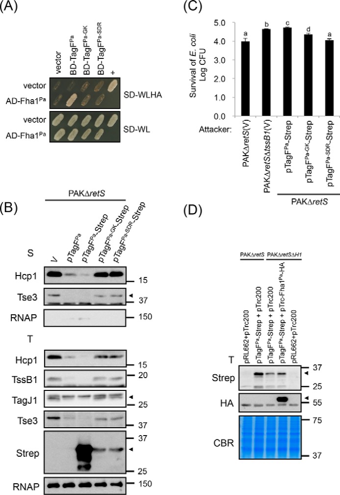
Conserved amino acid residues of TagFPa critical for TagFPa–Fha1Pa interaction are required for repressing H1-T6SS activity in P. aeruginosa. A, yeast two-hybrid protein–protein interaction results with P. aeruginosa Fha1 and various P. aeruginosa TagF proteins. SD−WL medium (SD minimal medium lacking Trp and Leu) was used for selecting plasmids. SD−WLHA medium (SD minimal medium lacking Trp, Leu, His, and Ade) was used for auxotrophic selection of bait and prey protein interactions. The positive interaction was determined by growth on SD−WLHA medium at 30 °C for at least 2 days. The positive control (+) showing interactions of SV40 large T-antigen and murine p53 and negative control (vector) are indicated. B, P. aeruginosa H1-T6SS secretion analysis. Shown is Western blot analysis of total (T) or secreted (S) proteins isolated from P. aeruginosa PAKΔretS (H1-T6SS–induced) harboring the vector pRL662 (V) or PAKΔretS harboring various overexpressed plasmids grown in TSB with specific antibodies. The nonsecreted RNA polymerase β subunit (RNAP) was an internal control. The proteins analyzed and molecular weight standards are shown on the left and right, respectively, and indicated with an arrowhead when necessary. C, P. aeruginosa H1-T6SS–mediated antibacterial assay against E. coli. Overnight cultures of P. aeruginosa PAKΔretS or PAKΔretSΔtssB1 (T6SS-defective strain) harboring the vector pRL662 (V) or various tagF-Strep–overexpressing plasmids were mixed with equivalent numbers of E. coli DH5α carrying a plasmid (pCR2.1) expressing β-gal. Data are mean ± S.D. of at least three biological replicates. Different letters above the bar indicate statistically significantly different groups of strains (p < 0.05) based on cfu of the surviving target cells. D, the presence of Fha1Pa increases the stability of TagFPa protein in P. aeruginosa. Shown is Western blot analysis of total (T) proteins isolated from P. aeruginosa PAKΔretS (H1-T6SS–induced) or PAKΔretSΔH1 (deletion of retS and H1-T6SS cluster) harboring various plasmid combinations grown in TSB with specific antibodies. All protein samples were analyzed by SDS-PAGE followed by Coomassie Blue staining (CBR) and served as an internal control. The proteins analyzed and molecular weight standards are shown on the left and right, respectively, and indicated with arrowheads when necessary.
