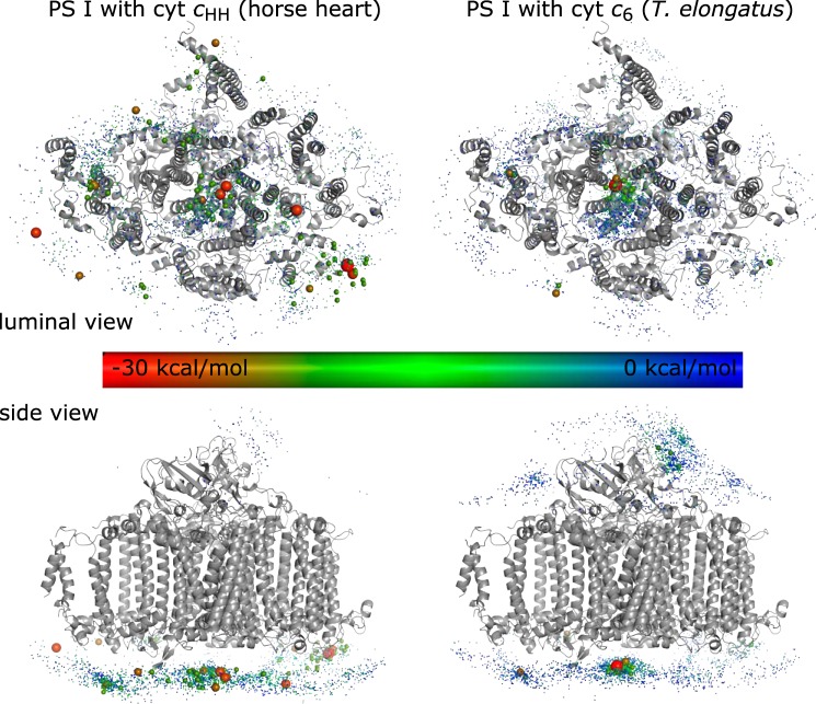
Figure 4.
Molecular docking simulation of monomeric PS I with cyt cHH (left) and cyt c6 (right). Each sphere represents the position of a docked cyt c. The binding energy, calculated by pyDock, is highlighted by a color code. Docking states with less than −20 kcal/mol are highlighted by an increased sphere size.

