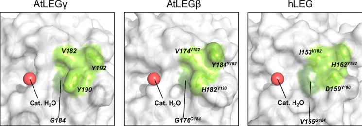
Figure 5.
S2′ pockets for different legumain isoforms and position of the catalytic water. Different legumain isoforms are displayed in surface representation. Bright and dark green are the wall- and bottom-forming residues of the different legumain S2′ pockets, respectively. A red sphere indicates the position of the catalytic water. The PDB ID of human legumain (hLEG) is 4AWA. The structure of AtLEGβ has been built as an homology model using AtLEGγ as a starting model. The homology model of the catalytic domain of AtLEGβ was built using the software Phyre2 (54).

