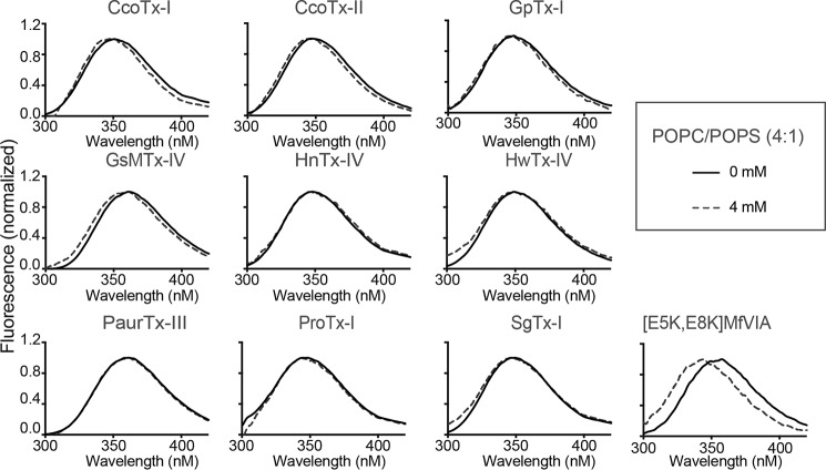
Figure 6.
Analysis of the environment around GMT Trp residues upon lipid titration. Fluorescence emission spectra of the peptides were followed in the absence (0 mm) and presence (4 mm) of POPC/POPS (4:1) LUVs. Excitation was at λ = 280 nm; peptide concentrations were 25 μm for all GMTs except PaurTx-III (12.5 μm) in HEPES-buffered saline. Spectra for [E5K,E8K]MfVIA, are included as a positive control (45).

