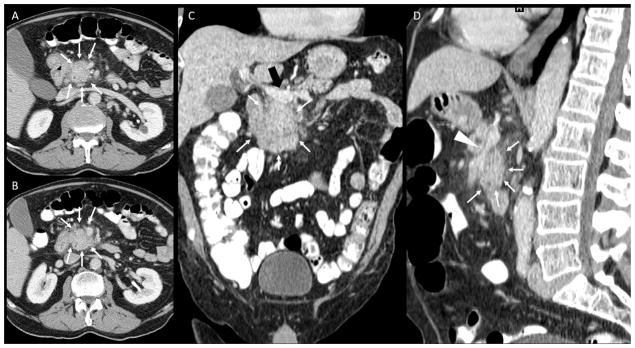
Figure 2.
Contrast CT of the abdomen, portal-venous phase. A, B: axial images; C: coronal reconstruction; D: Sagittal reconstruction. A pancreatic tumor is noted (white arrows in A, B, C, D) with peripancreatic fat infiltration, abuttment of portal vein at the spleno-mesenteric confluence (black arrow in C) and superior mesenteric vein encasement and narrowing (arrowhead in D). No significant artery involvement was described on outside report (T3, stage IIa). Second opinion report described the tumor encasement of the superior mesenteric artery (white arrowhead) changing thus the management of the patient (T4, stage III, unresectable).

