Fig. 8.
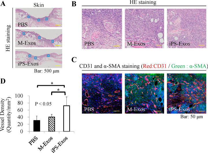
Immunofluorescence analyses of vessel formation
(A) Blood vessel numbers were determined in two areas at wound edges and three areas near the center of wounds.
(B) Representative hematoxylin & eosin staining images.
(C) Representative CD31 and α-SMA co-staining images, showing newly-formed vessels at wound sites.
(D) Quantification of average vessel density in each group. * P < 0.05.

