. 2018 May 31;25(2):123–132. doi: 10.11005/jbm.2018.25.2.123

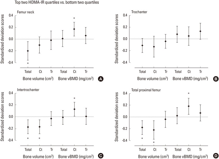
Fig. 2. Standardized difference (in standard deviation [SD] units±95% confidence interval) in quantitative computed tomography-derived bone volume and density at proximal femur for subjects in the top quartiles of homeostasis model assessment of insulin resistance (HOMA-IR) vs. lower quartiles after adjustment for age, weight, and height. Dashed line indicates the mean of the bottom two quartiles. The mean difference between the groups were expressed in SD scores as divided by the SD of the bottom two quartile group. Asterisk (*) indicated P<0.05 for the difference between the top and bottom two quartiles. vBMD, volumetric bone mineral density.

Fig. 2