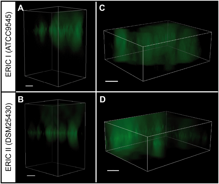
Figure 5.
Fluorescence microscopy of P. larvae floating biofilms. Bacterial suspensions of P. larvae ERIC I (ATCC9545; A,C) and P. larvae ERIC II (DSM25430; B,D) in Sf-900 II SFM medium supplemented with 30 µg/ml thioflavin S were incubated without agitation in 96-well-plates at 37 °C for six days. The thioflavin S-stained extracellular matrix in the floating biofilms was visualized using fluorescence microscopy; Z-stack processing was performed to obtain three-dimensional images of the wells containing the biofilms (A,B) and the region within the wells where the biofilms were located (C,D). Bars represent 20 µm.

