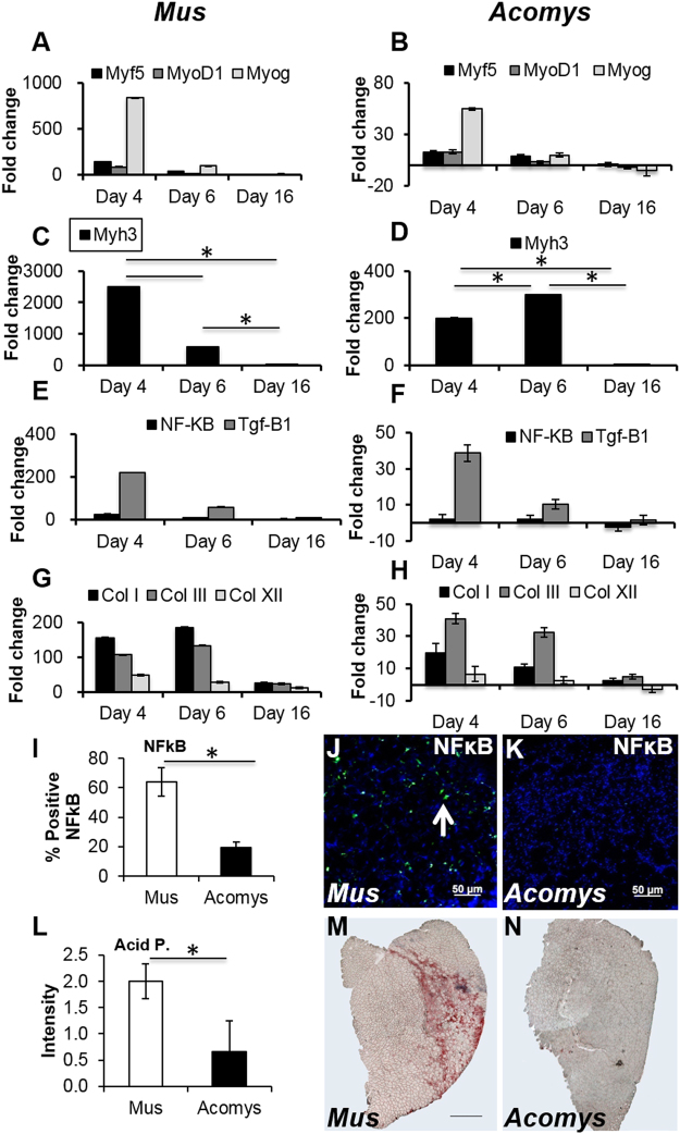
Figure 4.
Gene expression profiling, NFκB and lysosomal activity of CTX damaged skeletal muscle. Days 4, 6 and 16 after CTX injection in M. musculus (A,C,E,G) and A. cahirinus (B,D,F,H) TA. (A–B) Expression of Myf5, MyoD1 and Myogenin. (C–D) Expression of Myh3. (E–F) Expression of NFκB. (G–H) Expression of Collagen I, III and XII. (I) Quantification of NFκB positive cells in regenerating TA at day 6. (J–K) Immunocytochemical images for NFκB expressing in regenerating TA at day 6 in M. musculus and A. cahirinus (white arrow). (L) Quantification of Acid Phosphatase activity in regenerating TA at day 6. (M–N) Histological stained images for Acid Phosphatase activity in regenerating TA at day 6 in M. musculus and A. cahirinus. Statistical analysis performed by two-tailed t-test. *<0.05.

