Figure 7.
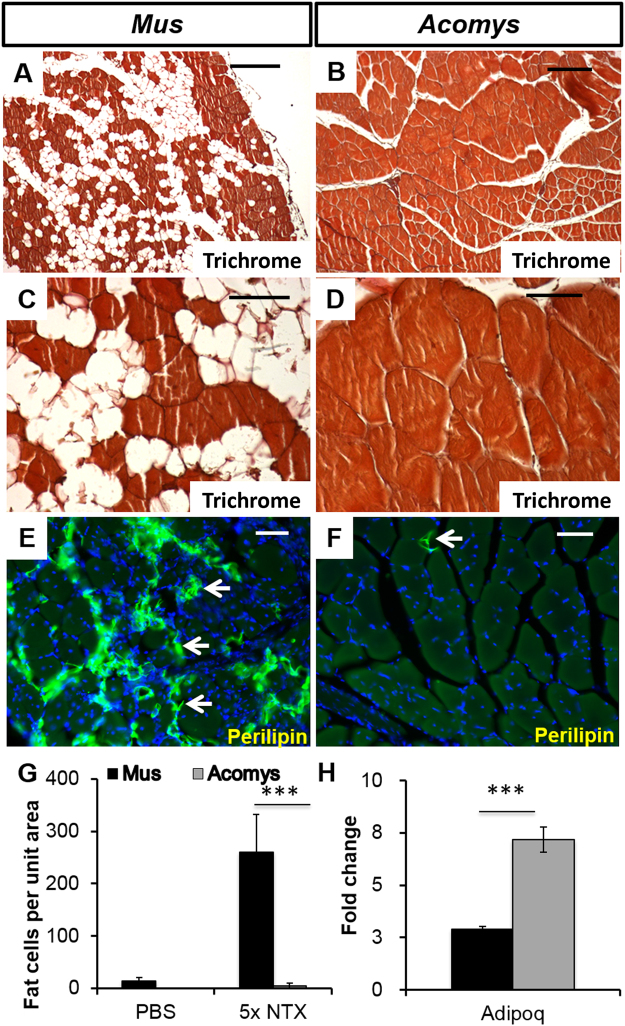
The effect of repeated rounds of cardiotoxin injury (5x) on muscle structure in M. musculus (left columns) and A. cahirinus (right columns). (A,C) Trichrome stained TA of M. musculus showing large gaps both within and between muscle fascicles. These are adipocytes (E). Scale bars = 100 μm (A) and 50 μm (C).(B,D) Trichrome stained TA of A. cahirinus showing perfect structural regeneration. Scale bars = 100 μm (B) and 50 μm (D). (E,F) Perilipin immunocytochemistry of M. musculus (E) showing many positive adipocytes (arrows) in the muscle compared to A. cahirinus (F) showing only one adipocyte present (arrow). Scale bars = 100 μm. (G) counts of adipocyte numbers in M. musculus and A. cahirinus TA after 5 rounds of repeated CTX injection into the TA showing almost no adipocytes present in A. cahirinus. (H) RT-qPCR analysis of gene expression for Adipoq after 5 rounds of CTX injections showing 2.5-fold higher levels in A. cahirinus (grey bar) which may contribute to its improved regenerative ability. Statistical analysis performed by two-tailed t-test. ***<0.001.

