Figure 1.
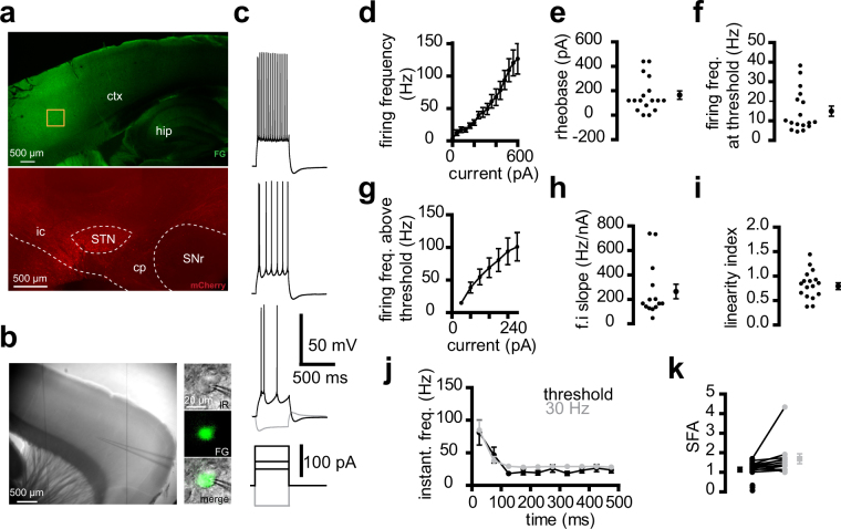
Properties of cortico-subthalamic neurons. (a) Confocal images of the cortex (top) after an injection of Fluoro-Gold (FG)retrograde tracer in the STN. To check the location of the injection site (bottom), the FG solution was mixed with a solution of anterograde virus expressing mCherry that labelled neurons at the injection site. FG and mCherry were detected by immunocytochemistry in a 350 μm slice used for electrophysiology. The orange box on the cortex indicates a high-density zone of FG-labelled neurons. Ctx, cortex; hip, hippocampus; ic, internal capsule; cp, cerebral peduncle; SNr, substantia nigra reticulata. (b) Infrared view of a living slice (left) and an FG-positive neuron at high magnification (right). (c) Example of the electrical properties of a retrograde-labelled cortico-subthalamic neuron. (d–i) Input-output relationship of a sample of 17 cortico-subthalamic neurons. (d) Mean firing frequency-current relationship. (e) Rheobase. (f) Firing frequency at threshold. (g) Mean firing frequency-current relationship above threshold. The current values represent the amplitude of step stimuli minus the amplitude of the first action potential-evoking step. (h) Firing frequency–current (f.i) slope. (i) Linearity index of the firing frequency–current relationship. e,f,g,h,i, left: individual values, right: mean ± sem. (j,k) Spike frequency over time in the same sample. (j) Instantaneous firing rate versus time (time was divided into 50 ms bins; times shown are the center of each bin) and (k) Spike firing adaptation, with black and grey symbols for values at threshold and 30 Hz, respectively. Three neurons with firing frequencies close to 30 Hz at threshold and those with above threshold values much higher than 30 Hz were not included in the 30 Hz data. (c–k) Saline was supplemented with 20 μM DNQX, 50 μM APV, 50 μM picrotoxin, and 1 μM CGP 55845.

